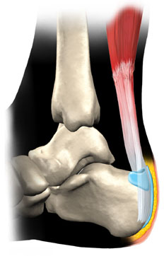
Mortons Neuroma Treatment

Overview

interdigital neuromaMorton?s neuroma occurs as the nerve passes under the ligament connecting the toe bones (metatarsals) in the forefoot. Morton?s neuroma most frequently develops between the third and fourth toes, usually in response to irritation, trauma or excessive pressure. The incidence of Morton?s neuroma is 8 to 10 times greater in women than in men.

Causes

A Morton’s neuroma commonly occurs due to repetitive weight bearing activity (such as walking or running) particularly when combined with tight fitting shoes or excessive pronation of the feet (i.e. “flat-feet”). The condition is also more common in patients with an unstable forefoot allowing excessive movement between the metatarsal bones. A Morton’s neuroma can also occur due to certain foot deformities, trauma to the foot, or the presence of a ganglion or inflamed bursa in the region which may place compressive forces on the nerve.

Symptoms

It usually occurs in between the 3rd and 4th toes (about 65% of cases) as is pictured to the right. It is less commonly found in the 2nd webspace, and rarely at all in the 1st or 4th webspaces. You can also experience pins and needles and/or numbness as a result of the nerve being affected. The condition tends to occur predominantly in middle aged females.

Diagnosis

To confirm the diagnosis, your doctor will examine your feet. He or she will look for areas of tenderness, swelling, calluses, numbness, muscle weakness and limited motion. To check for a Morton’s neuroma, your doctor will squeeze the sides of your foot. Squeezing should compress the neuroma and trigger your typical pain. In some cases, your doctor will find numbness in the webbed area between the affected toes. Pain in two or more locations on one foot, such as between both the second and third toes and the third and fourth toes, more likely indicates that the toe joints are inflamed rather than a Morton’ neuroma.

Non Surgical Treatment

Wear shoes with plenty of room for the toes to move, low heels, and laces or buckles that allow for width adjustment. Wear shoes with thick, shock-absorbent soles, as well as proper insoles that are designed to keep excessive pressure off of the foot. High-heeled shoes over two inches tall should be avoided whenever possible because they place undue strain on the forefoot. Resting the foot and massaging the affected area can temporarily alleviate neuroma pain. Use an ice pack to help to dull the pain and improve comfort. Use over-the-counter shoe pads. These pads can relieve pressure around the affected area.interdigital neuroma

Surgical Treatment

If conservative treatment does not relieve your symptoms, your orthopaedic surgeon may discuss surgical treatment options with you. Surgery can resect a small portion of the nerve or release the tissue around the nerve, and generally involves a short recovery period.

Tips On How To Diagnose Heel Spur

Inferior Calcaneal Spur

Overview

A common cause of heel pain is the heel spur, which is a bony growth on the underside of the heel bone. The spur, visible by X-ray, appears as a protrusion that can extend forward as much as half an inch. When there is no indication of bone enlargement, the condition is sometimes referred to as ?heel spur syndrome.?

Heel spurs result from strain on the muscles and ligaments of the foot, by stretching of the long band of tissue that connects the heel and the ball of the foot, and by repeated tearing away of the lining or membrane that covers the heel bone. These conditions may result from biomechanical imbalance, running or jogging, improperly fitted or excessively worn shoes, or obesity. Heel spurs specifically relate to the bony spurs (extra bony growth) that form at the base of the heel. It is a ?spike? of bone that grows from the base of the heel.

Causes

Heel spurs occur when calcium deposits build up on the underside of the heel bone, a process that usually occurs over a period of many months. Heel spurs are often caused by strains on foot muscles and ligaments, stretching of the plantar fascia, and repeated tearing of the membrane that covers the heel bone. Heel spurs are especially common among athletes whose activities include large amounts of running and jumping. Risk factors for heel spurs include walking gait abnormalities,which place excessive stress on the heel bone, ligaments, and nerves near the heel. Running or jogging, especially on hard surfaces. Poorly fitted or badly worn shoes, especially those lacking appropriate arch support. Excess weight and obesity. Other risk factors associated with plantar fasciitis include increasing age, which decreases plantar fascia flexibility and thins the heel’s protective fat pad. Diabetes. Spending most of the day on one’s feet. Frequent short bursts of physical activity. Having either flat feet or high arches.

Calcaneal Spur

Symptoms

Heel spur is characterised by a sharp pain under the heel when getting out of bed in the morning or getting up after sitting for a period of time. Walking around for a while often helps reduce the pain, turning it into a dull ache. However, sports, running or walking long distance makes the condition worse. In some cases swelling around the heel maybe present.

Diagnosis

Diagnosis of a heel spur can be done with an x-ray, which will be able to reveal the bony spur. Normally, it occurs where the plantar fascia connects to the heel bone. When the plantar fascia ligament is pulled excessively it begins to pull away from the heel bone. When this excessive pulling occurs, it causes the body to respond by depositing calcium in the injured area, resulting in the formation of the bone spur. The Plantar fascia ligament is a fibrous band of connective tissue running between the heel bone and the ball of the foot. This structure maintains the arch of the foot and distributes weight along the foot as we walk. However, due to the stress that this ligament must endure, it can easily become damaged which commonly occurs along with heel spurs.

Non Surgical Treatment

The first line of treatment for Heel Spur is to avoid the activities and positions that cause the pain. A physician can evaluate your foot with an X-ray to diagnose Heel Spur and determine a course of treatment. This condition can often be treated by non-surgical means; however in severe cases surgery may be necessary to relieve the pain. The most common surgical procedures treat the soft tissues around the Heel Spur, often a tarsal tunnel release or a plantar fascia release. Injections for heel spurs are sometimes controversial as steroids may cause heel pad atrophy or damage the plantar fascia.

Surgical Treatment

Sometimes bone spurs can be surgically removed or an operation to loosen the fascia, called a plantar fascia release can be performed. This surgery is about 80 percent effective in the small group of individuals who do not have relief with conservative treatment, but symptoms may return if preventative measures (wearing proper footwear, shoe inserts, stretching, etc) are not maintained.

Bursitis Foot Pain Symptoms

Overview

Bursitis and tendinitis are conditions that are also known as soft tissue rheumatic syndromes. A syndrome is a group of signs and symptoms that occur together and indicate a particular problem. This type of syndrome produces pain, swelling, or inflammation in the tissues and structures around a joint, such as the tendons, ligaments, bursae, and muscles.

Causes

Your ankle bursitis may have been caused by one or more of the following Pressure on your ankle and heels. This is often caused by running or exercising on uneven ground. The way that you exercise may also cause ankle bursitis or make it worse. It may be caused by wearing poorly fitting shoes that constantly rub against the heel. Direct, hard hit to your heel. Infection (in-FEK-shun). Medical problems such as rheumatoid (ROO-ma-toid) arthritis (ahr-THREYE-tis) or gout. Overusing your ankles. This is caused by doing activities or sports that use the same motions (movements) over and over again. Examples of repeating motions are running, walking, or jumping. Sometimes people do not know how they developed ankle bursitis.

Symptoms

Pain at the back of the heel at the attachment site of the Achilles tendon when running. Pain on palpation of the back of the heel or bottom of heel. Pain when standing on tiptoes. Swelling and redness at the back and bottom of the heel.

Diagnosis

When a patient has pain in a joint, a careful physical examination is needed to determine what type of movement is affected and if there is any swelling present. Bursitis will not show up on x-rays, although sometimes there are also calcium deposits in the joint that can be seen. Inserting a thin needle into the affected bursa and removing (aspirating) some of the synovial fluid for examination can confirm the diagnosis. In most cases, the fluid will not be clear. It can be tested for the presence of microorganisms, which would indicate an infection, and crystals, which could indicate gout. In instances where the diagnosis is difficult, a local anesthetic (a drug that numbs the area) is injected into the painful spot. If the discomfort stops temporarily, then bursitis is probably the correct diagnosis.

Non Surgical Treatment

The underlying cause of the bursitis must be identified to prevent further reoccurrences. Failure to eliminate the cause will lead to future flare ups and a poor and slow recovery. Future occurrences can be prevented with the use of stretches and strengthening exercises which will help prevent the muscles from tightening up over the bursa, Pilates and yoga are very good for this providing the aggravating movements are avoided. Wearing joint supports such as knee pads or elbow supports may also reduce the likelihood of redeveloping bursitis.

Surgical Treatment

Surgery is rarely need to treat most of these conditions. A patient with a soft tissue rheumatic syndrome may need surgery, however, if problems persist and other treatment methods do not help symptoms.

Prevention

Because many soft tissue conditions are caused by overuse, the best treatment is prevention. It is important to avoid or modify the activities that cause problems. Underlying conditions such as leg length differences, improper position or poor technique in sports or work must be corrected. Be aware of potential overuse or injury in your daily activities and change your lifestyle to prevent problems. Otherwise, problems may persist or occur repeatedly. Following are some ways you can avoid future problems. Wear walking or jogging shoes that provide good support. High-top shoes provide support for people with ankle problems. Wear comfortable shoes that fit properly. Wear heel cups or other shoe inserts as recommended by your doctor. Exercise on level, graded surfaces.

What Are Hammertoes

HammertoeOverview

A Hammer toes is a misshapen toe. The middle joint of the toe bends up in a way that makes the toe look like it is forming an upside-down V. The bent joint may rub the top of your shoe. Hammertoes can develop on any toe, but they usually happen in the second toe. Claw toes or mallet toes look a lot like hammertoes, but a different joint in the toe is bent. Hammertoes usually are not painful at first. When they begin, they can be pushed down to the correct position. These are called flexible hammertoes. After a while, they will not go back to their normal position, even if pushed with the fingers. These are called rigid hammertoes.

Causes

While most cases of hammertoes are caused by an underying muscle imbalance, it may develop as a result of several different causes, including arthritis, a hereditary condition, an injury, or ill-fitting shoes. In some cases, patients develop hammertoes after wearing shoes or stockings that are too tight for long periods of time. These patients usually develop hammertoes in both feet.

Hammer ToeSymptoms

Patients with hammer toe(s) may develop pain on the top of the toe(s), tip of the toe, and/or on the ball of the foot. Excessive pressure from shoes may result in the formation of a hardened portion of skin (corn or callus) on the knuckle and/or ball of the foot. Some people may not recognize that they have a hammer toe, rather they identity the excess skin build-up of a corn.The toe(s) may become irritated, red, warm, and/or swollen. The Hammer toe pain may be dull and mild or severe and sharp. Pain is often made worse by shoes, especially shoes that crowd the toes. While some hammer toes may result in significant pain, others may not be painful at all. Painful toes can prevent you from wearing stylish shoes.

Diagnosis

Your doctor is very likely to be able to diagnose your hammertoe simply by examining your foot. Even before that, he or she will probably ask about your family and personal medical history and evaluate your gait as you walk and the types of shoes you wear. You’ll be asked about your symptoms, when they started and when they occur. You may also be asked to flex your toe so that your doctor can get an idea of your range of motion. He or she may order x-rays in order to better define your deformity.

Non Surgical Treatment

If you have hammer toe, avoiding tight shoes and high heels may provide relief. Initial (non-surgical) treatment for hammer toe involves wearing shoes with plenty of room in the toe area. Shoes should be at least one-half inch longer than the longest toe. Stretching and strengthening exercises for the toes (such as picking up items with the toes or stretching the toes by hand) are also recommended. Sometimes orthopedists recommend special pads, cushions, or slings to help relieve the pain of hammer toe.

Surgical Treatment

Probably the most frequent procedure performed is one called a Post or an Arthroplasty. In this case a small piece of bone is removed from the joint to straighten the toe. The toe is shortened somewhat, but there is still motion within the toe post-operatively. In other cases, an Arthrodesis is performed. This involves fusing the abnormally-contracted joint. The Taylor procedure fuses only the first joint in the toe, whereas the Lambrinudi procedure fuses both joints within the toe. Toes which have had these procedures are usually perfectly straight, but they take longer to heal and don’t bend afterwards. A Hibbs procedure is a transfer of the toe’s long extensor tendon to the top of the metatarsal bone. The idea of this procedure is to remove the deforming cause of the hammertoes (in this case, extensor substitution), but to preserve the tendon’s function in dorsifexing the foot by reattaching it to the metatarsals. Fortunately, the Gotch (or Gotch and Kreuz) procedure–the removal of the base of the toe where it attaches to the foot, is done less frequently than in years past. The problem with this procedure is that it doesn’t address the problem at the level of the deformity, and it causes the toe to become destabilized, often resulting in a toe that has contracted up and back onto the top of the foot. You can even have an Implant Arthroplasty procedure, where a small, false joint is inserted into place. There are several other procedures, as well.

Overpronation Of The Feet

Overview

Over Pronation (Flat Feet) refers to the biomechanical shock-absorbing motion of the ankle, foot and lower leg. It is the natural inward flexing motion of the lower leg and ankle. Standing, walking, and running cause the ankle joint to pronate which in turn helps the body to absorb shock and allows it to control balance. An ankle joint that is too flexible causes more pronation than desired. This common condition is called Over- Pronation (sometimes referred to as “Flat Feet”). This foot condition places an extreme degree of strain on various connective tissues of the ankle, foot, and knee. If this condition is not addressed foot pain and toe deformities such as bunions and hammer toes (just to name a couple) may develop. Hip and lower back pain may also be residual results from this condition.Foot Pronation

Causes

Over-pronation has different causes. Obesity, pregnancy, age or repetitive pounding on a hard surface can weaken the arch leading to over-pronation. Over-pronation is also very common with athletes, especially runners and most of them nowadays use orthotics inside their shoes. Over-pronation affects millions of people and contributes to a range of common complaints including sore, aching feet, ball of foot pain, heel Pain, achilles tendonitis, bunions, shin pain, tired, aching legs, knee pain and lower back pain. The most effective treatment solution for over-pronation is wearing an orthotic shoe insert. Orthotics correct over-pronation, thereby providing natural, lasting pain relief from many common biomechanical complaints.

Symptoms

In addition to problems overpronation causes in the feet, it can also create issues in the calf muscles and lower legs. The calf muscles, which attach to the heel via the Achilles tendon, can become twisted and irritated as a result of the heel rolling excessively toward the midline of the body. Over time this can lead to inflexibility of the calf muscles and the Achilles tendon, which will likely lead to another common problem in the foot and ankle complex, the inability to dorsiflex. As such, overpronation is intrinsically linked to the inability to dorsiflex.

Diagnosis

A quick way to see if you over-pronate is to look for these signs. While standing straight with bare feet on the floor, look so see if the inside of your arch or sole touches the floor. Take a look at your hiking or running shoes; look for wear on the inside of the sole. Wet your feet and walk on a surface that will show the foot mark. If you have a neutral foot you should see your heel connected to the ball of your foot by a mark roughly half of width of your sole. If you over-pronate you will see greater than half and up to the full width of your sole.Foot Pronation

Non Surgical Treatment

Heel counters that make the heel of the shoe stronger to help resist/reduce excessive rearfoot motions. The heel counter is the hard piece in the back of the shoe that controls the foot?s motion from side-to-side. You can quickly test the effectiveness of a shoe?s heel counter by placing the shoe in the palm of your hand and putting your thumb in the mid-portion of the heel, trying to bend the back of the shoe. A heel counter that does not bend very much will provide superior motion control. Appropriate midsole density, the firmer the density, the more it will resist motion (important for a foot that overpronates or is pes planus), and the softer the density, the more it will shock absorb (important for a cavus foot with poor shock absorption) Wide base of support through the midfoot, to provide more support under a foot that is overpronated or the middle of the foot is collapsed inward.

Surgical Treatment

Depending on the severity of your condition, your surgeon may recommend one or more treatment options. Ultimately, however, it’s YOUR decision as to which makes the most sense to you. There are many resources available online and elsewhere for you to research the various options and make an informed decision.

What Would Cause Severs Disease?

Overview

When recurring heel pain occurs in children, it is usually due to Sever’s Disease, while adult heel pain is usually due to heel spurs, plantar fasciitis, or retrocalcaneal bursitis (Haglund’s Deformity). Calcaneus is the anatomical name of the heel bone. Sever’s Disease or Calcaneal Apophysitis is an inflammation of the growth plate located at the posterior aspect (back) of the heel.

Causes

The spontaneous development of pain in children generally indicates some form of injury to the growth plate of a growing bone. This can occur without a specific memorable event. When pain occurs in the heel of a child the most likely cause is due to injury of the growth plate in the heel bone. This is called Sever’s disease. A condition that may mimic Seiver’s disease is Achilles tendonitis. Achilles tendonitis is inflammation of the tendon attached to the back of the heel. A tight Achilles tendon may contribute to Sever’s disease by pulling excessively on the growth plate of the heel bone. It is frequently seen in the active soccer, football or baseball player. Sport shoes with cleats seem to aggravate the condition. It is believed that the condition is due to an underlying mechanical problem with the way the foot functions.

Symptoms

Patients with Severs disease typically experience pain that develops gradually in the back of the heel or Achilles region. In less severe cases, patients may only experience an ache or stiffness in the heel that increases with rest (especially at night or first thing in the morning). This typically occurs following activities which require strong or repetitive contraction of the calf muscles, such as running (especially uphill) or during sports involving running, jumping or hopping. The pain associated with this condition may also warm up with activity in the initial stages of the condition. As the condition progresses, patients may experience symptoms that increase during activity and affect performance. Pain may also increase when performing a calf stretch or heel raise (i.e. rising up onto tip toes). In severe cases, patients may walk with a limp, have difficulty putting their heel down, or be unable to weight bear on the affected leg. Pain may also increase on firmly touching the affected region and occasionally a bony lump may be palpable or visible at the back of the heel. This condition typically presents gradually overtime and can affect either one or both lower limbs.

Diagnosis

Physical examination varies depending on the severity and length of involvement. Bilateral involvement is present in approximately 60% of cases. Most patients experience pain with deep palpation at the Achilles insertion and pain when performing active toe raises. Forced dorsiflexion of the ankle also proves uncomfortable and is relieved with passive equinus positioning. Swelling may be present but usually is mild. In long-standing cases, the child may have calcaneal enlargement.

Non Surgical Treatment

The primary method of treating Sever?s disease is taking time off from sports and other physical activities to alleviate the pressure on the heel bone. During the healing period, your child?s doctor may also recommend physical therapy or any type of exercise that involves stretching and strengthen leg muscles and tendons. Wrapping ice in a towel and placing it under the child?s heel will also help to alleviate and reduce pain and swelling.

Prevention

It is important to undertake correct warm ups and warm downs before and after exercise. This should include a stretching routine. It may be necessary to undertake additional stretching outside of sport, especially during stages of growth. Only playing one sport should be avoided. You should not allow your child to play through pain.

Coping With Achilles Tendon Ruptures

Overview
Achilles Tendinitis
The Achilles tendon is situated above the heel and forms the lower part of the calf muscles. It is a continuation of the two calf muscles, the gastrocnemius and soleus muscles, and it attaches to the heel bone. It is the strongest tendon in the human body and must withstand great forces. Its function is to transmit the force produced by the calf muscles to lift the heel and produce the push off during walking, running and jumping. The Achilles can produce force of up to seven times body weight. This shows just how much force it has to withstand during sporting activities, such as sprinting, jumping and turning.

Causes
A rupture occurs when a patient overstretches the Achilles tendon, an act which causes it to tear partially or completely. Achilles tendon ruptures can occur during athletic play or any time the tendon is stretched in an unexpected way.

Symptoms
You may notice the symptoms come on suddenly during a sporting activity or injury. You might hear a snap or feel a sudden sharp pain when the tendon is torn. The sharp pain usually settles quickly, although there may be some aching at the back of the lower leg. After the injury, the usual symptoms are a flat-footed type of walk. You can walk and bear weight, but cannot push off the ground properly on the side where the tendon is ruptured. Inability to stand on tiptoe. If the tendon is completely torn, you may feel a gap just above the back of the heel. However, if there is bruising then the swelling may disguise the gap. If you suspect an Achilles tendon rupture, it is best to see a doctor urgently, because the tendon heals better if treated sooner rather than later. A person with a ruptured Achilles tendon may experience one or more of the following. Sudden pain (which feels like a kick or a stab) in the back of the ankle or calf, often subsiding into a dull ache. A popping or snapping sensation. Swelling on the back of the leg between the heel and the calf. Difficulty walking (especially upstairs or uphill) and difficulty rising up on the toes.

Diagnosis
The diagnosis is usually made on the basis of symptoms, the history of the injury and a doctor’s examination. The doctor may look at your walking and observe whether you can stand on tiptoe. She/he may test the tendon using a method called Thompson’s test (also known as the calf squeeze test). In this test, you will be asked to lie face down on the examination bench and to bend your knee. The doctor will gently squeeze the calf muscles at the back of your leg, and observe how the ankle moves. If the Achilles tendon is OK, the calf squeeze will make the foot point briefly away from the leg (a movement called plantar flexion). This is quite an accurate test for Achilles tendon rupture. If the diagnosis is uncertain, an ultrasound or MRI scan may help. An Achilles tendon rupture is sometimes difficult to diagnose and can be missed on first assessment. It is important for both doctors and patients to be aware of this and to look carefully for an Achilles tendon rupture if it is suspected.

Non Surgical Treatment
The best treatment for a ruptured Achilles tendon often depends on your age, activity level and the severity of your injury. In general, younger and more active people often choose surgery to repair a completely ruptured Achilles tendon while older people are more likely to opt for nonsurgical treatment. Recent studies, however, have shown fairly equal effectiveness of both operative and nonoperative management. Nonsurgical treatment. This approach typically involves wearing a cast or walking boot with wedges to elevate your heel; this allows the ends of your torn tendon to heal. This method can be effective, and it avoids the risks, such as infection, associated with surgery. However, the likelihood of re-rupture may be higher with a nonsurgical approach, and recovery can take longer. If re-rupture occurs, surgical repair may be more difficult.
Achilles Tendon

Surgical Treatment
An Achilles tendon rupture is a complete tear of the fibrous tissue that connects the heel to the calf muscle. This is often caused by a sudden movement that overextends the tendon and usually occurs while running or playing sports such as basketball or racquetball. Achilles tendon rupture can affect anyone, but occurs most often in middle-aged men.